Saturday, November 17, 2007

How Long To Do Zumba Before Seeing Effects


biological tissue is supportive, with function together other tissues and structures.
It originates from the mesoderm.
is the one that serves to unite, support and protect body parts .

Features:

Their cells are embedded and widely dispersed in an abundant intercellular material, called the extracellular matrix.

represents the extravascular, interstitial space agency .


Structure:

Cells:

own or fixed cells

Responsible for the formation and maintenance

tissue they belong to, because

by what are called

supporting cells

  • own or fixed cells.
  • free conjunctival cells

Matrix extracellular

  • fundamental substance.

  • conjunctive fibers.


own or fixed cells:

Responsible for the formation and maintenance of the tissue to which they belong, which is why they are called supporting cells.

They can be distinguished as

  • mesenchymal cells . Fibroblasts
  • and fibrocytes
  • reticular cells
  • fat cells.

mesenchymal cells : are precursor cells. They are located in the vessel walls.

Fibroblasts and fibrocytes:
are responsible for:
- production collagen fibers , reticular and elastic.

- production glycosaminoglycans and glycoproteins of fundamental amorphous substance .-

These substances produce involved in wound healing and tissue restoration.


fibroblasts.

Reticulum cells:

produce reticular fibers.


fat cells:

is a specialized cell in the storage of neutral fats.


free conjunctival cells or wandering:

  • mast cells

  • macrophages.

  • plasma cells.

  • lymphocytes.

  • granulocytes polymorphonuclear .


mast cells or mast cells :

precursors are derived from the bone marrow .

participate in the inflammatory response of the immune system .

mast cells or mast cells.


Macrophages:

are large cells in the form of stars.

remove foreign substances and are involved in immune response.


Macrophages


plasma cells:

Soon many in normal tissue.
participate in the processes of chronic inflammation .

Its function is to synthesize the antibodies found in blood.


plasma cells.
Other tissue cells blood derivatives:
  • white blood cells.
  • Lymphocyte.
  • Eosinophils.


fibers.

  • collagen.

  • Waffle.
  • Elastic
  • .
collagen fibers:

They are most abundant in connective tissue. are constituted by a scleroprotein called collagen.


fibers Waffle:

collagen fibers are very thin.

networks is presented as a way around fibers muscle, nerve fibers, fat cells and small blood vessels.

also in smaller divisions lung and particularly, within the limits between connective tissue and other tissue types.


Elastic Fibers:

are thinner than the fibers of elastic fibers easily give colágeno.Las the minimum traction and can be stretched.

The main component is the protein elastin.

Found in: arterial walls, bronchi, bronchioles, ligaments yellow ligaments vocalesy spine.


fundamental substance.

is clear, colorless and homogeneous.

occupies the spaces between cells and connective tissue fibers.

is formed mainly by: glycosaminoglycans and glycoproteins .


- Connective Tissue Types and location:


- Connective tissue mucous. Is forming pulp cartilaginous intervertebral discs.

-areolar or loose connective tissue. Located as part of the dermis of the skin and in the stroma of the organs like the liver, spleen, etc.

- dense connective tissue. Located as part of some layers of the skin) dermis and epidermis).

- Connective tissue fat. It is found mainly in the subcutaneous tissue.

- reticular connective tissue. As part of the stroma of organs like the spleen.

- elastic connective tissue. Located in the walls of blood vessels as large arteries in ligaments and the vocal cords.

- Connective tissue bone and cartilage. Forms the skeleton and joints.


mucosal connective tissue:

Appears in the normal development and differentiation of connective tissues.

is the main component umbilical cord. An Appeal Wharton jelly .

is found in the nucleus pulposus of intervertebral discs and dental pulp.

forms an elastic cushion to protect against the pressure surrounding structures.


- loose connective tissue or areolar:

has abundant ground substance.

Rich in cells of various tipos.Fibras collagen and elastic thin and sparse. Fill the spaces between the fibers and make musculares.Se in the skin, mucous membranes and glands in .

loose connective tissue is delicate consistency, flexible and very resistant to traction.


dense connective tissue:

predominant collagen fibers . The cells are escasas.Se tissue is less flexible and much more resistant to traction.

Classified as:

regular dense connective tissue.
irregular dense connective tissue.

regular dense connective tissue:

Contains fibers and grouped close together and parallel to each other to form structures with high resistance to stress. is found in ligaments, joints and wrap a few wraps of the organs.

- Function:
- transmit mechanical strength to the muscles.
- Join the bones together.
- forms the protective covering (capsule) of organs.


irregular dense connective tissue:

is presented in the form of hojas.Las fibers interlock to form a mesh-resistant.

is located in the dermis and most organs wrappers.

Its functions:

- Resist the pressure from all directions .

- Protects fragile bodies.


reticular connective tissue:
- Consisting reticular fibers in close association with primitive reticular cells.

is mainly found in the bone marrow, spleen and lymph nodes.

Their function is

- Hold for cell phones.

- important in filtering the blood.


elastic connective tissue:

Formed by parallel thick elastic fibers. Presents yellow and has remarkable elasticity . Form
yellow ligament vertebral column ligament and suspensory penis.
p is found in Aredes of the great arteries, vocal chords trachea, bronchi.


connective tissue adipose

variety of loose connective tissue. Fat cells are numerous and very pressed together. has a small amount of elastic fibers, reticular and collagen .

- Function:
-
Mechanics: fill gaps and reduce the effect pressures.
- Power reserve.


connective tissue functions:


- serves as a vehicle for the vessels, nerves and excretory ducts . Transportation.

- Has mechanical functions Hold and barrier .

- healing and repair of tissues

- defending the body against harmful infectious agents or other

- function storage . water reserve and electrolytes.
- spoke on the immune system. Immune Defense.

- Nutrition: transport serves nutritional substances that will lás cells.

Spy Camara Brazilian Wax

tissues. Epithelial tissue.

tissue is called to the group of cells with a specific structure to perform a specialized function, vital for the body.

Group of cells, fibers and cell products forming a set number of structural and fulfilling the same function.


Rating:


According to its nature and function

differ in:

1 .- epithelial tissue.

  • epithelial tissue lining.
  • glandular epithelial tissue.
  • sensory epithelial tissue.

2 .- Connective tissue.

  • connective tissue itself.
  • specialized connective tissue:
    • bone tissue.
    • cartilage.
    • blood tissue.

3 .- muscle tissue.

  • striated muscle.
  • smooth muscle.
  • heart muscle.
4 .- Nervous tissue.

Epithelial Tissue:

The epithelium is the tissue formed by one or more layers of cells juxtaposed to form the inner lining of the cavities, hollow organs, ducts of the body and skin, and also are the mucous membranes and glands.

is that generally consists of polyhedral cells, juxtaposed, among which there is little intercellular substance.


Features:

consists of cells closely set together. With little or no intercellular substance. The cells are arranged in a single layer or multiple layers.


Structure:

- Cells:

  • flat cells
  • cubic cells
  • cylindrical cells.


Union means:

  • - Union Cement
  • - desmosomes
  • - Bands closing
  • - Unions close
  • - interdigitating membranes
  • - membrane folds
  • - Membrane basal


Polarity:

Each cell has three types of surfaces apical surface (free) a number variable side surfaces adjacent to neighboring cells and a basal surface attached to the basal layer.


Specializations epithelial cells:

apical surface specializations:
- cilia: the airway epithelium and genital .

striated ( microvilli ): intestinal epithelium.


brush border: renal tubular epithelium.


embryonic epithelial tissue Source:

, originating from the three layers germ. Ectoderm, endoderm and mesoderm.


Ectoderm:


are derived from this:
  • surface of the skin epithelium.
  • sensory epithelium (nasal mucosa, retina, inner ear, taste buds).

Mesoderm:

are derived from this:

  • epithelium of the seminiferous tubules in the testes and ovaries germinal epithelium.
  • Endothelium of the heart and blood vessels.
  • pericardium (membrane that surrounds the heart), pleura (membrane covering the lungs and inside the chest) and peritoneum (lining of the abdominal organs and inside the abdomen).
  • brain membranes (meninges), inner ear, eye and genital tract.

Endoderm:

  • Epithelium of the digestive tract and accessory glands (liver, pancreas).
  • epithelium of the airways and lungs.
epithelia Rating:

Depending on the function of the epithelium:

  • - lining epithelium or squamous
  • - lar glandular epithelium.
Skin Glands

Depending on the shape of epithelial cells:

  • - squamous epithelium or squamous. Squamous cells are flat and similar to plates.
  • - cuboidal epithelium. cuboid cells are polygonal with

    height and width similar.

  • - Epithelium prismatic or cylindrical. cylindrical cells are polygonal with greater height than width.


Depending on the number of layers of cells that form:

  • - simple epithelium. A single layer.
  • - stratified epithelium. Two or more cell layers.


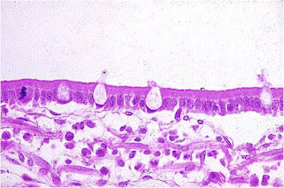
• Epithelium pseudostratified :
are characterized by:
All the cells rest on the sheet baseline.
• Not all extend to the surface.
nuclei are at different depths .


epithelial tissue:

specific epithelial types.

simple squamous epithelium:
single layer of flat cells
• They act as a semipermeable barrier between compartments.
• Location:
Blood vessels (endothelium).
body cavities (mesothelium ).

renal glomeruli.


cuboidal epithelium:
single layer of cuboidal cells or in a brick .
• Form the walls of the ducts.
• Carries secretion and excretion products.
• Location:
• kidney tubules.
small gland ducts.
• Crystal (capsule).


Epitelio cilíndrico simple:
Capa única de células aproximadamente

cilíndricas.

Superficies apicales (libres) pueden estar

cubiertas de cilios o microvellosidades .

Join secretion, absorption and

propulsion of mucus.

is a protective barrier.

Location:
• Stomach.
• small and large intestines.
• Uterus.
• Fallopian tube.
Conductos grandes glandulares. Algunos conductos renales (papilares).


Epitelio cilíndrico seudoestratificado.
Capa única de células de forma y altura variable.
Los núcleos se ubican en diferentes niveles.
cells often reach the surface are hair.


• Forms a protective barrier.
• Mobilization of mucus with cilia.
• Location:
• Epithelial airways.
• Male reproductive system.

stratified squamous epithelium:
can be:
• stratified squamous epithelium keratinized :
• Multiple layers of cells.
surface cells are squamous dead and filled with keratin. Without core.
• Deeper layers have cells polygonal in progressive stages of keratinization .


• Location:
Mainly in the skin.
highly specialized forms a barrier against friction abrasion infection and water loss.

• stratified squamous epithelium not keratinized :
is thinner.
surface cells are flattened nucleated.
is also called mucosa.
• Forms a protective barrier less resistant.
Location: mouth, vagina, esophagus, anal canal, vocal cords.


cubic stratified epithelium:
has two to three layers of cuboidal cells.
is rare and scarce.
• Location:
• Of ducts some glands (salivary, sweat).


epithelium cylindrical stratified:
• Similar to the cubic layered.
Its cells are cylindrical surface and may have cilia.
• Location:
• Of the ducts over

some large glands.

• Conjunctiva. (Eye membrane)
respiratory surface of the epiglottis.


E pitelio Transitional:
• Also known as mixed or polymorphic.
is considered as a variety of epithelial layered cylindrical.
so named because the cells that make up undergo changes in shape and position, as the state of relaxation or contraction of the organ

lining.

• Forms the lining of most of the tracks urinary tract (renal pelvis, ureters, bladder, etc..).


glandular epithelial tissue:


Glands:

are single cells or groups of cells specialized for secretion. All glands arise in early development, from the lining or covering epithelium.


glandular epithelial tissue:

is characterized by the property they own their cells produce substances that pass then to the outside or the circulation, to be distributed throughout the body.

• Depending on the number of cells that compose it classified as:

  • unicellular glands.
    • Sometimes, among the cells lining epithelium isolated glandular cells interspersed.


  • multicellular gland. entire lining epithelium is at once secretory


Glands:

can be of two types according to where

Pour

their secretions

• exocrine glands , are retaining their connection with the epithelium through a conduit.

• Endocrine Glands (ductless glands) lose their connections with the surface and release their secretions into the bloodstream.


Category

GLANDS

EXOCRINE

GLANDS

ENDOCRINE

Liberation

secretions

Through

ducts

A circulation

Number

cells

Unicelares or

multicellular

multicellular

secretory products

Protein

glycoproteins and

some lipids

peptide hormones and

hormones. steroid


function of epithelia:

  • Injury Protection. Covering (epidermis)
  • discharge of substances (glands)
  • substance absorption (intestine, kidney)
  • sensory reception (sense organ)
  • sensory function. Excretion
  • (Kidney)
  • Transportation (lung)
  • digestion (digestive tract)