Monday, November 19, 2007

Muvies En Espana En Espanol Gratis

Bone and cartilage tissue.

tissues are characterized because their cells are surrounded by an intercellular matrix solid and relatively rigid .

cartilage.
has rigid consistency. Its surface is slightly elastic and very smooth.
is composed of a rich array

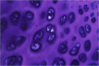
the extracellular which the chondrocytes (cartilage cells) are located in spaces called lacunae.


Functions:

  • it supports or support.
  • Of the articular surfaces.
  • is part of the airways.


Structure:


cells.

-chondroblasts.
-Chondrocytes.

Matrix extracellular. It consists of:
  • • Collagen.
    • Elastin.
    hyaluronic acid
    • Glycoproteins
    • Proteoglycans


Chondrocytes synthesize and secrete organic components of the extracellular matrix. The death of the cells leads to degeneration

matrix. blood vessels do not penetrate the cartilage matrix
. They feed material broadcasting from the tissue capillaries adjacent .

is surrounded by the perichondrium corresponding to dense connective tissue.

Types:

According with the abundance and type of fiber in the matrix:


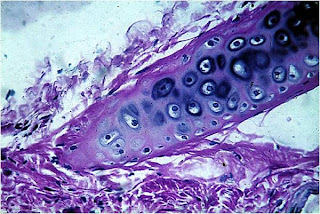
• hyaline cartilage:
  • is the most common.
  • has a moderate amount of collagen fibers .
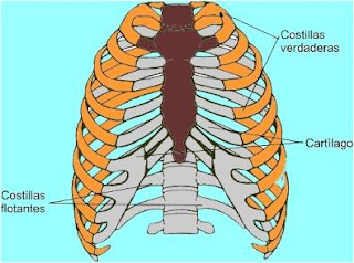
  • forms the fetal skeleton, epiphyseal plates , joints, c. sack and large airways.


• Elastic cartilage:
structure similar to hyaline cartilage.
has collagen fibers and abundant elastin fibers.
is located in the outer ear, trunk Eustáquio , epiglottis, cartilage corniculate and cuneiform of the larynx.


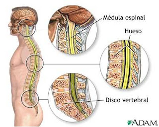
• Fibrous Cartilage:
The matrix consists almost entirely fibers collagen forming a network.

• Found in intervertebral discs, symphysis pubis.
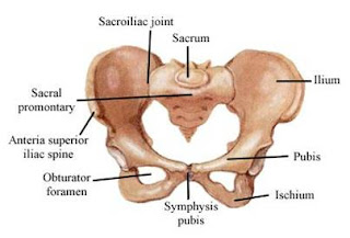


bone tissue.


Bone tissue is a type of connective tissue is characterized by its rigidity and its resistance both tensile and compressive .
is a tough and resilient tissue formed by cells bone called "osteoblasts."


Structure:

Trained by:
• Bone matrix: intercellular calcified material.
• Cells:
  • Osteoblasts: responsible to produce and secrete the organic part of bone matrix during its formation. Located at the periphery of the tissue.
  • osteocytes are responsible for the maintenance of bone matrix. They are located in cavities or gaps.
  • Osteoclasts, cells responsible for resorption of bone tissue located in Howship gaps.
• The bone matrix is \u200b\u200bdeposited inorganic salts granted hardness. These are:
calcium phosphate.
• calcium carbonate.
• Calcium fluoride.
magnesium fluoride.

bone Rating:


• spongy bone:
Formed by thin
trabeculae. Presents bone marrow-filled cavities.
• Function: involved in hematopoiesis (blood formation).
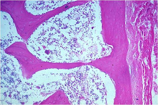
is found in flat bones and ends of long bones .
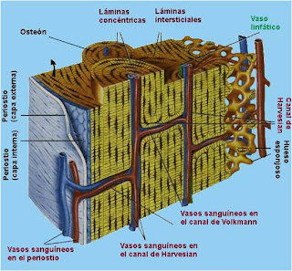

• Compact bone:
Formed by plates or concentric rings around central channel called Havers channels. channels Havers are connected to other channels channels called Volkmann. Among the concentric lamellae of mineralized matrix there are small holes or gaps where are the osteocytes.

is located in the outer layer of flat bones and bodies of long bones.


functions of bone tissue:

• Form the skeleton that supports the body.
constitute the insertion point of muscles .
• Contains the bone marrow where blood cells are formed.


Use Motorcycle Engine In Go Kart


The nervous tissue is scattered by the body interrelate and forming a communication network is the nervous system.

is composed of neurons that are specialized cells in the electrochemical conduction of nerve impulses.


Features:

  • It originates from the ectoderm.
  • Its main components are cells surrounded by scarce intercellular material.

  • is located at the Central Nervous System and Peripheral Nervous System .


Structure:

is formed by:

- Cells

  • neurons or nerve cells.

  • Neuroglia or supporting cells.


nerve cells or neurons.

are the functional cells of nervous tissue. Are interconnected to form networks.


Functions:

receive and integrate stimuli simultaneously from various sources.
transmit these nerve impulses.


Structure:

  • The cell body includes the cytoplasm and nucleus.
  • The dendrites. The function of dendrites is to receive stimuli from the environment, epitelialessensoriales cells or other neurons.
  • The

    axon or axis cylinder. The axon is the transmitter with respect

    the nerve impulse, or transmitting the nerve impulse. This has the following structures.

    • myelin sheath.
    • cells Shwann .
    • Ranvier nodules.


Synapse: The

synapses (the gr. σύναψις, "link") are specialized junctions through which cells nervous system send signals to each other and non-neuronal cells such as muscle or glandular .

A synapse between a motor neuron and muscle cell called neuromuscular junction.

synapses allow neurons the central nervous system form a network of neural circuits.

In a prototypical synapse, such as those found in dendritic buttons , Cytoplasmic projections with mushroom from each cell, and in which both ends are crushed against each other. In this area, cell membranes of both cells were close together in a union that allows signaling molecules called neurotransmitters quickly switch to another cell by diffusion . The canal linking the postsynaptic neuron, is about 20 nm wide, and is known as synaptic cleft.


synapses are made between the axon of one neuron and the dendrites or the body of other neurons.

Classification of neurons:

According to the number of extensions.

  • Unipolar or seudomonopolares .


  • Bipolar.


  • Multipole .


According to its function.

  • motor neuron. Carry

    nerve impulse to the organs peripheral terminals.

  • sensory neuron.
    receive momentum generated by stimulation and transported to the central nervous system.
  • intermediate neuron.
    interconnect two neurons: a motor with a sensitive, sensitive to sensory, motor to another.

supporting cells.


surround the neurons and play support functions, defense, nutrition and adjusting the composition of intercellular material.


Nerve Fibers.

is formed by the axon and its associated sheaths.
The axon from its beginning to near its termination is surrounded by a sheath Shwann . older axons inside the sheath of Shwann are surrounded by a myelin sheath. In Central Nervous System can be covered in part by glial cells ( unmyelinated) or have a pod

myelin formed by oligodendrocytes ( Melina).


nerve fibers are composed by grouping a large number of axons and these in turn are combined to form the nerves.


Nervios.

Formados por agrupaciones de fibras nerviosas en forma de haces en el Sistema Nervioso Periférico .
Poseen fibras aferentes y eferentes.
  • Las aferentes llevan a los centros las informaciones procedentes del organismo y del medio ambiente.
  • Las fibras eferentes transmiten impulsos de los centros nerviosos a los órganos efectores controlled by these centers.
  • nerves may be sensory, motor and mostly mixed.

functions of nervous tissue:

  • from collects information from sensory receptors .
  • process this information, providing memory system.

  • generates proper signals to

    Effector cells

Pain In Left Shoulder From Cold Drink

Nerve Tissue Connective Tissue Muscle tissue

Muscle tissue is responsible for the body movements. It consists of elongated cells called muscle fibers, characterized by presence of large numbers of cytoplasmic filaments.


is characterized by its contractility. is formed by elongated cells, also called muscle fibers. It originates from the mesoderm.

muscle fibers are limited outside a membrane called the sarcolemma. the cytoplasm called sarcoplasm is striated by the presence of myofibrils.


We can say that muscle tissue is a group or complex histological, because in addition to the type of muscle cells also have a tissue component conjunctive.


There are two types of myofibrils:
• Smooth, homogeneous structure.
• Ridged: heterogeneous for the presence of disks light and dark alternate regularly.

myofibril Depending on the make up muscle fiber can be:

• smooth muscle fiber.


• striated muscle fiber.


• Muscle fiber heart


muscle Classification:
According to their morphological and functional is classified as

Striated or skeletal muscle:

• Formed by bundles of cylindrical cells very long and multinucleate presenting transverse striations.
have fast twitch, vigorous and subject to Voluntary Control.


• Smooth muscle:
• Formed by clusters of spindle cells have no cross striations.
• Your contractions are slow and are not subject voluntary control.


cardiac striated muscle:
• Formed by elongated and branched cells.
These are attached longitudinally to neighboring forming a network.
• Your contractions are vigorous, involuntary and rhythmic.


striated muscle tissue or skeletal muscle:

Formed by striated muscle fibers, which are cells cilindroides , unbranched, longer than 1 to 4 cms of length. With many nuclei peripherals.


Organization Musculoskeletal:

The bundles of muscle fibers are wrapped in a membrane called epimysium .
• From epimisio start dividing walls to beams in installments and this partition is called perimysium .
Each muscle fiber is enveloped by a layer of reticular fibers called endomysium .
penetrate the muscle capillaries through connective septa.


flattened nuclei and peripherals are under the sarcolemma (plasma membrane of the muscle cell).
• cytoplasm or sarcoplasm area around the nuclei and myofibrils.

All of this together in structures called
Leydig cylinders or columns of Kölliker.


skeletal muscle tissue myofibrils and filaments have many heterogeneous.
Most of the organelles and sarcoplasm (cytoplasm) are close to the poles of the nuclei.
The sarcoplasm contains many mitochondria or sarcosomas , glycogen granules and a protein called oxygen-binding myoglobin .
The endoplasmic reticulum in these cells is called sarcoplasmic reticulum . It consists of a membrane system that extends throughout the muscle fiber and is attached at some sites to the sarcolemma. is related to the propagation of the wave of excitement that causes contraction of the fiber.


filaments in cells or muscle fibers may be:
-thin filaments.
Actin .
• Tropomyosin .
• Troponin .

-Fiber Thick:
• Myosin .


These filaments form the Myofibrils that are cylindrical structures that are distributed longitudinally occupying almost the entire interior muscle fiber.

• Myofibrils are arranged parallel to the axis fiber that contains them.
• They consist of two substances that in the form of records regularly called alternative bands.

  • Banda or disk A: dark filaments formed by myosin . This presented in its middle a zone clear close call Striae of or disk Hensen H.


  • Banda or disk I: filaments composed of actin . It surpasses in part and interdigitates with strands of myosin without the band H. This presented in this medium

    a dark area called Disco Z.


All these structures together in structural and functional unit called muscle: Sarcomere .


Striated Muscle Tissue Cardiac

formed by spindle cells, forming columns that irregularly anastomose or join. Unlike skeletal fibers have only one or two core areas. The direction of cardiac fibers is very irregular . The columns are covered by a thin sheath of connective tissue, equivalent to endomysium of skeletal muscle.


heart muscle fiber is very similar to skeletal but has more sarcoplasm, mitochondria and glycogen.

Myofibrils are not arranged in bundles, are distributed throughout the sarcoplasm .

At the junction between two fibers is a line dark cross called intercalated disks and represent specialized areas for the union.


cardiac muscle fiber has the same bands A, I, H and Z of the skeleton but not the filaments are A bundle but form a large mass.

The sarcoplasmic reticulum is less developed in these cells compared with that of skeletal muscle fibers.

smooth muscle:


This association consists of large cells and spindle layered above in walls of hollow organs (intestines, vessels).

• smooth muscle Other locations are:
• Connective tissue surrounding:
• Prostate.
• seminal vesicles.
• The subcutaneous tissue:
• Scrotum.
Nipples.
• erector muscle of hair.
• Uterus.


smooth muscle fibers are coated and bonded together by a network of reticular fibers The smooth muscle cell is also coated externally by a layer glycoprotein amorphous ( glycocalyx).

has been shown that smooth muscle cells also contractile capacity, can also synthesize collagen type III fibers elastic and proteoglycans.


functions of muscle tissue.
Participate in all body movements
  • Volunteers (skeletal muscle)
  • Involuntary (smooth muscle, cardiac striated muscle).